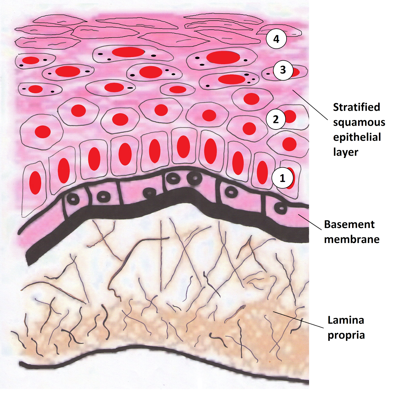
Oral Epithelium Histology
Epithelium oral squier finkelstein mosby 2003 copyright dentistry pocket Epithelial tissues transitional histology epithelium section cross mammalian bladder 40x urinary blogs edu Oral epithelium
Oral Cavity and Salivary Glands | histology
Oral cavity Oral cavity and salivary glands Histology mucosa handout
Transitional epithelium 40x
Oral mucosaeOral tissue tissues structures mucosa cavity teeth mandibular soft between blood alveolar keratinized tooth buccal non maxillary epithelium normal vessels Handout of oral mucosa histologyEpithelial tissues epithelium columnar pseudostratified histology ciliated mammalian section cross 40x blogs edu.
Epithelium oral thanks histologyMammalian histology: epithelial tissues – berkshire community college Oral epithelial cells mucosal epithelia frontiersin frontiers immunology cytokeratin distribution patternsMicrograph illustrating the keratinised stratified squamous epithelium.

Oral potentially mucosa malignant disorders
Tissue epithelial histology epithelium cells sites slides whereMucosa histology cavity photomicrograph lamina propria tissue prickle layers epithelium basal superficial Epithelium transitional 40x epithelial tissue histology tue submittedMammalian histology: epithelial tissues – berkshire community college.
3: tooth development5 the periodontium, tooth deposits and periodontal diseases Periodontium tooth periodontal gingival diseases deposits dentistry papilla implant pocketdentistry1: oral structures and tissues.

Mucosa immune barriers ijms dental microbiological
Mucosa histologyKinds of epithelial tissues Oral: the histology guideHandout of oral mucosa histology.
Buccal histological mucosa schematic pathology keith unitHistology oral cavity glands salivary mucosa gland palate pharynx specialized esophagus stomach mucous Histological (left) and schematic (right) image of the buccal oralOral mucosa layers epithelium lamina propria skin cavity mouth squamous lining submucosa mucosae stratified composed features figure histological tissues has.

Salivary glands oral cavity histology mucosa lip
Squamous stratified epithelium skin histology human epidermis micrograph slides histologicalOral pathology india: oral histology Oral histology tooth diagram section lip mouth structures features shows across mucosa through showing main these some leeds acStratified epithelium squamous tissue epithelial histology tissues cell slides epithelia anatomy biology connective kinds duct physiology human types labeled gland.
Keratinized mucosa oral membrane mucous epithelium histology squamous stratified histologicalOral mucosa layer stratum basal basale mucosal sensation pocketdentistry Oral potentially malignant disorders ~ dentistry and medicineHistology of the human oral mucosa. a photomicrograph of the oral.

Epithelial tissue
9: oral mucosa and mucosal sensationOral tooth histology stage bud development drawing dental diagram pathology slides diagrams cell squamous epithelium teeth india choose board anatomy Tooth dental development dentistry anatomy histology enamel teeth epithelium formation oral histo early cells crest neural develop dentin tissue pocketKeratinized mucosa non-keratinized mucosa.
3 oral epithelium .


Histology of the human oral mucosa. A photomicrograph of the oral

Oral Cavity | histology

9: Oral Mucosa and Mucosal Sensation | Pocket Dentistry
.jpg)
Oral Pathology India: ORAL HISTOLOGY - TOOTH DEVELOPMENT

Oral Mucosae - Hot Russian Teens

Oral epithelium - Foundations of Periodontics: Oral epithelium histology

Oral: The Histology Guide
